BOLD signal
BOLD fMRI is often used to measure the hemodynamic response resulting from changes in the oxygenation level in the blood vessels. Yet to be explained is the relationship between neuronal activity and the measured fMRI signal. In a seminal paper, Ogawa et al., (1990) showed that blood-oxygenation-level-dependent (BOLD) constitutes a natural contrast for fMRI that can be used to image brain function.
The BOLD signal does not directly reflect spiking activity of neurons, rather it is a metabolic-related signal that accompanies changes in neuronal activity. If BOLD increases in a certain brain region then it can be said that it reflects the processing of information by a local neuronal activity (including excitatory and inhibitory interneurons).
Currently it is thought that BOLD mainly correlates with vascular density and the number of synapses, and less with spiking activity of neurons (Logothetis and Pfeuffer, 2004). However, in response to external stimuli, both neuronal synaptic and spiking activity consume energy in the form of glucose that is supplied back through the blood stream. Energy consumption can also be expressed in terms of oxygen consumption (oxygen cerebral metabolic rate, CMRO2) that covaries with neuronal activity. Additionally, neuronal activity causes increased blood flow to the activated region (increased cerebral blood flow, CBF) and expansion of the blood vessels (increased cerebral blood volume, CBV). Oxygen is carried by the hemoglobin in red blood cells, which leads to a local oxygenated blood increase, with simultaneous decrease in deoxygenated blood.
While diamagnetic oxyhemoglobin blood has no interference with the magnetic field, paramagnetic deoxyhemoglobin induces local field inhomogeneities, reducing the BOLD signal. This magnetic property difference is what allows the identification and localization of the activated region. In fact, BOLD fMRI measures exactly the changes in local magnetic field inhomogeneities using the aforementioned EPI sequences (see here) sensitive to these susceptibility differences (Logothetis and Pfeuffer, 2004).
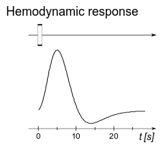
The BOLD fMRI signal is characterized by the hemodynamic response function (HRF), assumed to be the response of the system to a brief and intense period of neural activity. The nature of the BOLD response determines the temporal sensitivity of fMRI. The HRF lasts for about 10 seconds (peaking at about 5 s) after stimulation (Figure 1).
References
1. Ogawa, S., Lee, T.-M., Nayak, A. S. & Glynn, P. Oxygenation-sensitive contrast in magnetic resonance image of rodent brain at high magnetic fields. Magn. Reson. Med. 14, 68–78 (1990).
2. Logothetis, N. K. & Pfeuffer, J. On the nature of the BOLD fMRI contrast mechanism. Magnetic Resonance Imaging 22, 1517–1531 (2004).
Note: In fMRI , spatial resolution depends on various scanning parameters such as the field strength or the coil adequacy to the structure being imaged. Additionally, spatial sensitivity is also determined by the size of the voxel (an element on a three dimensional space grid) that it is used, which depending on the study can range from 1 mm to 3 mm. Depending on the area being scanned and its size, one voxel can contain a few million neurons and tens of billions of synapses. A single voxel’s response signal over time is called its time course.
License

This work is licensed under a Creative Commons Attribution-NonCommercial 4.0 International License.
This article is part of the Introductory Chapter (Chapter 1) of A. Amaral’s doctoral thesis Functional MRI of stimulus interference on auditory processing in normal listeners and tinnitus patients available @ http://hdl.handle.net/10362/14989